Aromatase Is Ruining Your Life
Every morning you wake up and your body hands you testosterone.
If it turns into DHT, you get:
Energy.
Confidence.
Focus.
Drive.
Power.
If it turns into estrogen through aromatase, you get:
Tired.
Soft.
Foggy.
Flat.
Fat.
Same molecule. Same morning. One enzyme decides which way it goes.
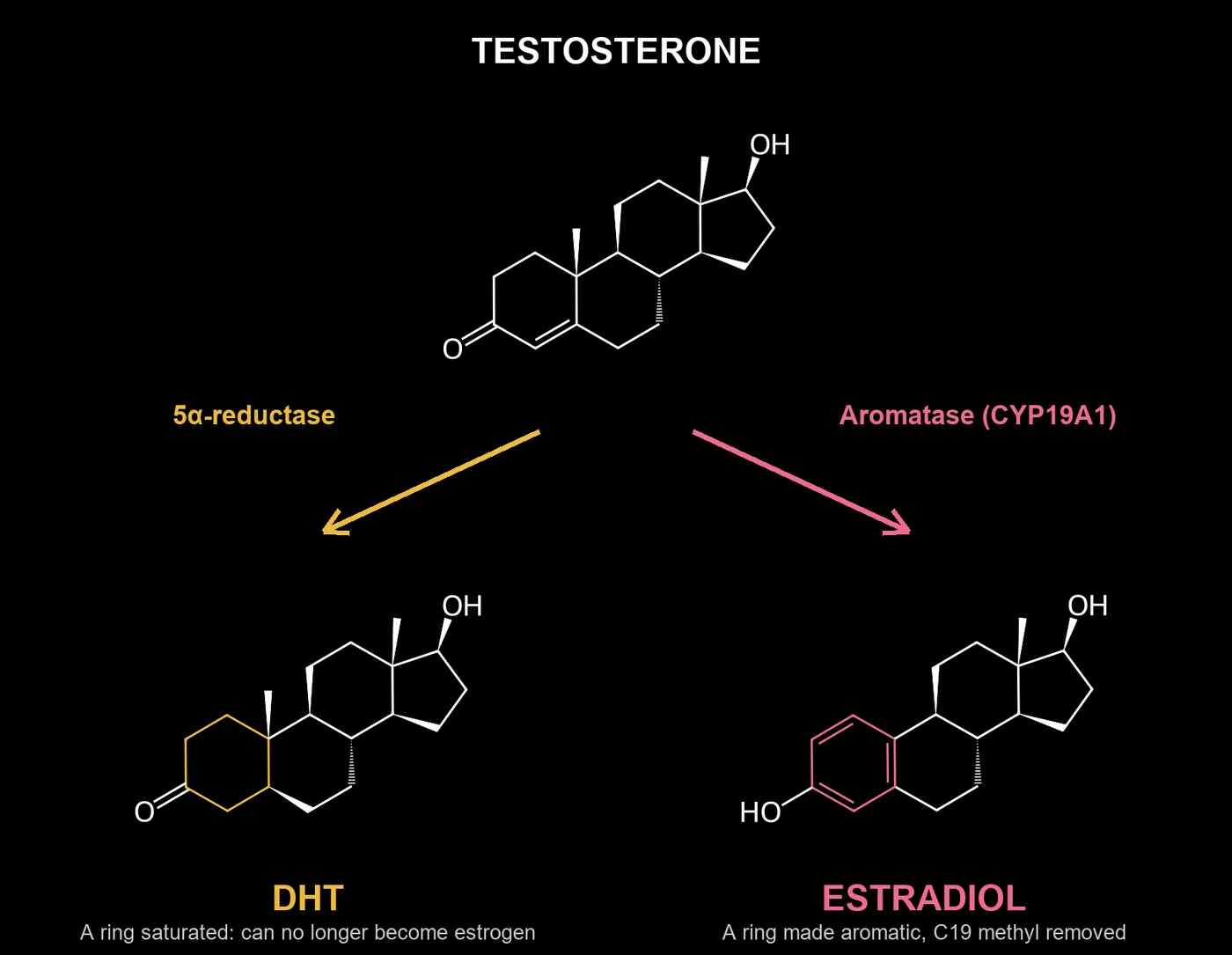
That enzyme is aromatase, the product of one gene, CYP19A1.
It lives in your fat, brain, bone, skin, liver and testes, and how much of it you run depends on how you live.
Below: 10 angles, 50 questions, 150 studies.
Every number on this page comes from the study's abstract on PubMed. Where the evidence is weak or points the other way, it says so.
1. The enzyme itself: what aromatase is and where it lives
1.1 CYP19A1: the one gene and enzyme that turns testosterone into estradiol and androstenedione into estrone
CYP19A1 is the gene that codes for aromatase, a cytochrome P450 enzyme sitting in the cell's endoplasmic reticulum.
It strips one carbon from an androgen and turns its first ring aromatic, which is the only chemical route the body has to make estrogen.
Testosterone becomes estradiol, androstenedione becomes estrone.
Every estrogen molecule in a man's body passed through this one door.
PMID 12053085 · The aromatase cytochrome P-450 and its clinical impact (2002)
Review: aromatase (P450arom) is encoded by a single gene, CYP19, on 15q21.2 (about 123 kb) and converts testosterone to estradiol and androstenedione to estrone; the protein is the same in every tissue.
PMID 19707181 · Aromatase deficiency in men: a clinical perspective (2009)
Clinical review of men with aromatase deficiency: loss-of-function mutations in CYP19A1 leave serum estradiol undetectable, with tall stature, unfused epiphyses and osteopenia or osteoporosis.
PMID 8076586 · Aromatase cytochrome P450, the enzyme responsible for estrogen biosynthesis (1994)
Classic Endocrine Reviews paper (Simpson et al.) describing aromatase cytochrome P450 as the enzyme responsible for estrogen biosynthesis; no abstract on PubMed, so no numbers.
1.2 Where it is found: fat, brain, bone, skin, liver, testes, breast, blood vessels
Aromatase is not confined to the ovaries.
It is active in fat, brain, bone, skin, liver, blood vessels, breast tissue and the testes.
That means every one of these tissues can make its own estrogen from circulating androgens.
The bigger and more active these tissues are, the more of your testosterone gets turned into estrogen on site.
PMID 15083376 · Organization of the human aromatase p450 (CYP19) gene (2004)
Review of the human CYP19 gene: its partly tissue-specific promoters are used in the gonads, bone, brain, vascular tissue, adipose tissue, skin, fetal liver and placenta to make estrogen.
PMID 11850209 · Aromatase in atherosclerotic lesions of human aorta (2001)
Human aorta from 85 autopsy cases: aromatase and 17beta-HSD I were found in smooth muscle cells of the vessel wall, especially next to atheromatous plaque, so estrone and estradiol are made in the artery wall.
PMID 16019206 · Aromatase in testis: expression and role in male reproduction (2005)
Testis review: in man, Leydig cells, immature germ cells and ejaculated spermatozoa express a biologically active aromatase (a single 49 kDa band on western blots).
1.3 Tissue-specific promoters: the same gene is switched on by different signals in different tissues
The CYP19A1 gene has several separate on-switches (promoters), one or more per tissue.
Fat uses a promoter driven by cortisol and cytokines, the gonads and breast tumors use one driven by cAMP and PGE2, and the placenta and brain have their own. This is why stress and inflammation can raise estrogen in belly fat while the testes or ovaries stay unchanged.
PMID 8476746 · Tissue-specific promoters regulate aromatase cytochrome P450 expression (1993)
Simpson lab: tissue-specific promoters drive aromatase in ovary (proximal promoter), placenta (promoter at least 40 kb upstream) and adipose tissue (adipose-specific promoter), yet the protein made is identical.
PMID 15083377 · Aromatase: biologic relevance of tissue-specific expression (2004)
Review: each aromatase promoter is regulated by a distinct signaling pathway in a tissue- and hormone-specific manner, covering ovary, adipose tissue, placenta, bone, brain and malignant cells.
PMID 9705080 · Aromatase expression of human osteoblast-like cells (1998)
Human osteoblast-like cells: aromatase was stimulated by dexamethasone plus cytokines (IL-1beta, TNFalpha, type I cytokines) and the major promoter used was I.4, unlike other estrogen-source tissues.
1.4 Local versus blood estrogen: tissue levels can be high while blood tests look normal
Most estrogen made outside the gonads acts inside the tissue that made it and never reaches the blood in large amounts.
Breast tumors, fat and the prostate can hold estrogen levels far above what a blood test shows.
A normal serum estradiol does not prove that a tissue is not flooded with estrogen.
PMID 9618794 · Concentration of sex steroids in adipose tissue after menopause (1998)
35 postmenopausal women without cancer and 19 breast cancer patients: mean sex steroid levels were higher in fat than in plasma, apart from testosterone.
PMID 24081738 · 17β-Estradiol and estradiol fatty acyl esters and estrogen-converting enzyme expression in adipose tissue in obese men and women (2013)
14 severely obese men and 22 premenopausal women at bariatric surgery: serum estradiol did not significantly correlate with estradiol in adipose tissue, so fat estrogen production was not reflected in blood.
PMID 14623515 · Sources of estrogen and their importance (2003)
Simpson review: in men and postmenopausal women estradiol is made and acts locally, so circulating levels reflect rather than direct estrogen action; breast tumor estrogen can be at least one order of magnitude above blood.
1.5 Men versus women: why the enzyme matters at every age in both sexes
In men, almost all estradiol comes from aromatase working on testosterone, much of it outside the testes.
In women, the ovaries dominate until menopause, after which fat and other tissues take over.
In both sexes, aromatase sets how much of the androgen supply ends up as estrogen, and that share tends to climb with fat and age.
PMID 16480891 · Of mice and men: the evolving phenotype of aromatase deficiency (2006)
Review of the seven known men with nonfunctional aromatase (CYP19 mutations): undetectable estrogen, normal to high testosterone, osteoporosis, impaired lipid and insulin metabolism and impaired reproductive function.
PMID 11826265 · Aromatase--a brief overview (2002)
Simpson review: in postmenopausal women, as in men, estrogen is made in breast, bone, vasculature and brain and acts locally; androgen precursors decline with age in women, a possible reason for more bone loss than in men.
PMID 28539434 · Estrogens in Male Physiology (2017)
Physiological Reviews: aromatase is expressed in Leydig cells, seminiferous epithelium and other male organs, and loss of ESR1 or aromatase affects brain, adipose, muscle, bone, cardiovascular and immune tissue in males.
2. Fat as an estrogen factory (the obesity loop)
2.1 Fat stromal cells as the main source of aromatase outside the gonads
The cells that make estrogen in fat are not the fat cells themselves but the stromal cells around them (preadipocytes and fibroblasts).
After menopause and in men, this tissue is the main estrogen source. More fat mass means more stromal cells, and more stromal cells means more aromatase.
PMID 7251819 · Aromatization of androstenedione by human adipose tissue stromal cells in monolayer culture (1981)
Adipose tissue from women: 87% of aromatase activity was in the stromal/vascular fraction and only 13% in adipocytes, so fat estrogen comes mainly from stromal cells.
PMID 6310250 · Aromatization of androgens by human adipose tissue in vitro (1983)
Cultured human adipose stromal cells: aromatase activity was stimulated 20-100-fold by dexamethasone and up to 200-fold by dibutyryl cyclic AMP after 8 days.
PMID 11511861 · Estrogen production and action (2001)
Review (Nelson and Bulun): aromatase in adipose tissue and possibly skin primarily accounts for extraglandular estrogen formation and increases as a function of body weight and advancing age.
2.2 The cycle: more fat → more aromatase → more estrogen → lower testosterone → more fat
Fat makes aromatase, aromatase turns testosterone into estrogen, and estrogen signals the brain to cut testosterone production.
Lower testosterone and higher estrogen favor fat storage, especially in the belly.
The loop is a strong hypothesis, but the one direct test is mixed: blocking aromatase in obese men raised testosterone without changing body fat.
PMID 10342671 · The hypogonadal-obesity cycle: role of aromatase in modulating the testosterone-estradiol shunt--a major factor in the genesis of morbid obesity (1999)
Hypothesis paper (Cohen) describing the hypogonadal-obesity cycle: more abdominal fat, more aromatase, more testosterone converted to estradiol, lower testosterone, more abdominal fat.
PMID 28679145 · The Effect of Aromatase on the Reproductive Function of Obese Males (2017)
Review: aromatase expression is proportional to body fat mass and causes more fat accumulation, a vicious cycle that lowers testosterone in obese men.
PMID 23949882 · Somatic and psychological effects of low-dose aromatase inhibition in men with obesity-related hypogonadotropic hypotestosteronemia (2013)
Direct test in 42 obese men (RCT): letrozole raised testosterone from 8.6 to 21.5 nmol/l and cut estradiol from 119.1 to 59.2 pmol/l, but had no significant effect on body composition or other endpoints.
2.3 Belly fat versus fat under the skin: does location change aromatase activity?
Not all fat behaves the same.
Belly (visceral) fat and fat under the skin differ in blood flow, inflammation and how aromatase is switched on.
Researchers have measured aromatase in both depots, and the results conflict: in men belly fat tracks estradiol, yet some tissue studies find more aromatase in fat under the skin.
PMID 22469460 · Associations between visceral adipose tissue, inflammation and sex steroid concentrations in men (2013)
229 dysmetabolic men (MRI in 109): visceral fat correlated with estradiol (r = 0.38) and inversely with the bioavailable testosterone/estradiol ratio (r = -0.42); subcutaneous fat correlated with neither.
PMID 12198740 · Estradiol in elderly men (2002)
Vermeulen review: in men estradiol relates to subcutaneous abdominal fat but not visceral fat, and aromatase activity in omental fat is only one-tenth of that in gluteal fat.
PMID 29029113 · Estrogen Metabolism in Abdominal Subcutaneous and Visceral Adipose Tissue in Postmenopausal Women (2017)
37 postmenopausal women: estrone was higher in visceral fat, but subcutaneous fat made more estradiol from estrone (1.02 vs 0.57 nmol/kg AT/h); visceral estradiol production rose with waist circumference.
2.4 Low testosterone in obese men driven by estrogen feedback on the pituitary (hypogonadotropic hypogonadism)
In obese men the testes are often healthy; the signal from the brain is what drops.
Estradiol made in fat feeds back on the hypothalamus and pituitary and lowers LH, so the testes are told to make less testosterone. This is called hypogonadotropic hypogonadism, and blocking aromatase can reverse part of it.
PMID 15811136 · Letrozole normalizes serum testosterone in severely obese men with hypogonadotropic hypogonadism (2005)
10 severely obese men with hypogonadotropic hypogonadism given letrozole for 6 weeks: estradiol fell from 120 to 70 pmol/l, LH rose from 4.5 to 14.8 U/l and testosterone rose from 7.5 to 23.8 nmol/l.
PMID 18426834 · Letrozole once a week normalizes serum testosterone in obesity-related male hypogonadism (2008)
12 severely obese men with hypogonadotropic hypogonadism on letrozole 2.5 mg once a week: estradiol fell from 123 to 58 pmol/l, LH rose from 4.4 to 11.1 U/l and testosterone rose from 5.9 to 19.6 nmol/l.
PMID 10999781 · Aromatase inhibition in the human male reveals a hypothalamic site of estrogen feedback (2000)
Normal and GnRH-treated hypogonadal men given anastrozole: estradiol fell (136 to 52 pmol/L in normal men) and testosterone rose about 53%, showing estradiol feedback at the hypothalamus as well as the pituitary.
2.5 Weight-loss studies: does testosterone come back as aromatase falls?
If fat drives aromatase, losing fat should reverse the picture.
Studies of diet and bariatric surgery in obese men track testosterone, estradiol and SHBG before and after weight loss.
They test whether testosterone recovers in proportion to the fat that is lost.
PMID 23482592 · Body weight loss reverts obesity-associated hypogonadotropic hypogonadism: a systematic review and meta-analysis (2013)
Meta-analysis of 24 studies: diet and bariatric surgery raised testosterone, lowered estradiol, and the amount of weight lost was the best determinant of the testosterone rise.
PMID 26892583 · Effect of Lifestyle Intervention on the Hormonal Profile of Frail, Obese Older Men (2016)
RCT in 40 frail obese men aged 65 or older: diet-induced weight loss of about 10 kg lowered total estradiol and raised total testosterone over 12 months; free testosterone index did not change significantly.
PMID 32700180 · Bariatric Surgery Impact on Reproductive Hormones, Semen Analysis, and Sperm DNA Fragmentation in Men with Severe Obesity: Prospective Study (2020)
Prospective study of men with severe obesity: obese men had higher estradiol and lower testosterone than controls, and after bariatric surgery total testosterone more than doubled (294.5 to 604 ng/dL).
3. Aging: the slow drift toward estrogen
3.1 Does aromatase activity in fat rise with age?
Aging changes fat tissue: more inflammation, more stromal cells and a shift in how the aromatase gene is regulated.
Studies have measured aromatase activity in fat and skin cells across age groups.
The question is whether the older body converts a larger share of its androgens into estrogen.
PMID 3964790 · Effects of aging and obesity on aromatase activity of human adipose cells (1985)
Adipose stromal cells from 50 women: the aromatization rate per cell increased with age (r = 0.43), while obesity raised estrogen output through more fat cells rather than higher activity per cell.
PMID 8989235 · Alternatively spliced transcripts of the aromatase cytochrome P450 (CYP19) gene in adipose tissue of women (1997)
Adipose tissue from the abdomen, buttocks and thighs of women: aromatase transcript levels increased in direct proportion to advancing age, highest in buttocks and lowest in abdomen.
PMID 4815174 · Plasma precursors of estrogen. II. Correlation of the extent of conversion of plasma androstenedione to estrone with age (1974)
Classic JCEM tracer study (Hemsell, MacDonald) correlating the conversion of plasma androstenedione to estrone with age; no abstract on PubMed, so no numbers.
3.2 Testosterone-to-estradiol ratio across a man's life
Testosterone falls slowly with age while estradiol in men stays flat or falls much less.
The result is a falling testosterone-to-estradiol ratio. Large cohort studies have tracked both hormones in men across decades and linked the ratio to fat, health and symptoms.
PMID 9554416 · Sex hormones and age: a cross-sectional study of testosterone and estradiol and their bioavailable fractions in community-dwelling men (1998)
Rancho Bernardo, 810 men aged 24-90: bioavailable testosterone and bioavailable estradiol both decreased with age independent of covariates.
PMID 15305335 · Serum sex hormones and measures of benign prostatic hyperplasia (2004)
Olmsted County men: bioavailable testosterone fell with age while the estradiol/bioavailable testosterone ratio rose from 0.042 to 0.044 to 0.050 across men under 60, 60-69 and over 69.
PMID 19896534 · Aromatase and regulating the estrogen:androgen ratio in the prostate gland (2010)
Review: serum testosterone drops by about 35% between ages 21 and 85 while estradiol stays constant or rises, and this changing T:E ratio is implicated in prostate disease.
3.3 After menopause, fat becomes the main estrogen source
When the ovaries stop, estrogen does not disappear.
It is made in fat, skin and other tissues from adrenal androgens through aromatase.
This is why a heavier postmenopausal woman has higher estrogen levels and why aromatase inhibitors work in postmenopausal breast cancer.
PMID 4688315 · Source of estrogen production in postmenopausal women (1973)
Classic JCEM study (Grodin, Siiteri, MacDonald) on the source of estrogen production in postmenopausal women; no abstract on PubMed, so no numbers.
PMID 10851574 · Aromatase in aging women (1999)
Review (Bulun, Simpson): after the menopause, aromatase in adipose tissue and possibly skin is the main source of estrogen, and it rises with body weight and advancing age.
PMID 24923731 · All sex steroids are made intracellularly in peripheral tissues by the mechanisms of intracrinology after menopause (2015)
Labrie review: after menopause all estrogens and androgens are made locally in peripheral tissues (intracrinology) and acted on there, with serum estradiol kept at very low levels.
3.4 Muscle loss, belly fat and "andropause" through the estrogen lens
Many signs blamed on andropause (belly fat, softer muscle, low drive) overlap with a shift toward estrogen.
Studies separate the effects of low testosterone from those of estradiol by giving testosterone while blocking its conversion.
The surprise: in the key trial, too little estrogen, not too much, drove the gain in body fat.
PMID 24024838 · Gonadal steroids and body composition, strength, and sexual function in men (2013)
NEJM RCT, 198 plus 202 healthy men aged 20-50 with gonadal suppression, testosterone gel with or without anastrozole: androgen deficiency drove loss of lean mass and strength, estrogen deficiency drove the rise in body fat.
PMID 26588809 · Effects of aromatase inhibition vs. testosterone in older men with low testosterone: randomized-controlled trial (2016)
RCT in 43 men aged 65-82 with low testosterone: anastrozole and testosterone gel both raised testosterone; lean mass rose only with anastrozole (1.49 kg at 6 months), but bone density improved only with testosterone.
PMID 11399122 · Aromatase, adiposity, aging and disease. The hypogonadal-metabolic-atherogenic-disease and aging connection (2001)
Hypothesis paper (Cohen): with aging, rising fat mass brings more aromatase, which converts testosterone to estradiol, lowers testosterone and favors visceral fat, feeding insulin resistance and muscle loss.
3.5 Prostate: the estrogen/androgen balance and BPH in older men
The aging prostate sits in a hormonal environment where androgens fall and estrogen holds steady.
Prostate stromal tissue expresses aromatase and estrogen receptors, and animal studies show that estrogen can drive prostate growth and inflammation. But in the direct human trial, an aromatase inhibitor did no better than placebo for established BPH.
PMID 1722796 · Rationale for using aromatase inhibitors to manage benign prostatic hyperplasia. Experimental studies (1991)
Review of experimental studies: rising BPH with age coincides with a shift in the androgen:estrogen ratio toward estrogens in serum and prostate, and aromatase inhibitors blocked androgen-driven stromal growth in dogs and monkeys.
PMID 21620560 · Androgens and estrogens in benign prostatic hyperplasia: past, present and future (2011)
Review: testosterone is converted by CYP19/aromatase to estradiol, the prostate is an estrogen target, and estrogen receptor signaling may drive BPH and urinary symptoms, but the evidence is still incomplete.
PMID 8876703 · Estrogen reduction by aromatase inhibition for benign prostatic hyperplasia: results of a double-blind, placebo-controlled, randomized clinical trial using two doses of the aromatase-inhibitor atamestane. Atamestane Study Group (1996)
Direct test, RCT in 292 men with BPH symptoms for 48 weeks: the aromatase inhibitor atamestane lowered estradiol and estrone but did no better than placebo for established BPH.
4. Stress, inflammation and the aromatase switch
4.1 Cortisol and glucocorticoids switching on aromatase in fat
Cortisol and synthetic glucocorticoids act on the promoter that fat uses for aromatase, and in cell studies they raise aromatase sharply, especially together with cytokines.
But in human fat tissue the direction depends on sex: cortisol raised aromatase in women's fat and lowered it in men's.
The stress-to-estrogen link is real in the lab, less simple in men.
PMID 11889205 · Glucocorticoid regulation of p450 aromatase activity in human adipose tissue: gender and site differences (2002)
In human preadipocytes from surgery patients, cortisol 10(-6) M raised subcutaneous aromatase activity in women from 11.5 +/- 1.4 to 28.0 +/- 1.8 pmol/mg x h but inhibited it in men (19.4 +/- 2.4 vs 7.5 +/- 1.3), while in omental fat cortisol raised activity in men from 2.6 +/- 0.1 to 7.8 +/- 0.3 pmol/mg x h.
PMID 7776980 · Characterization of the sequences of the human CYP19 (aromatase) gene that mediate regulation by glucocorticoids in adipose stromal cells and fetal hepatocytes (1995)
In human adipose stromal cells and fetal hepatocytes, dexamethasone induced CYP19 promoter I.4 reporter activity about 3-fold (-560/+10 construct) and 8-fold (-330/+170 construct containing a glucocorticoid response element), and mutating the GRE abolished the induction.
PMID 7608217 · Aromatase P450 gene expression in human adipose tissue. Role of a Jak/STAT pathway in regulation of the adipose-specific promoter (1995)
In human adipose stromal cells the adipose aromatase promoter I.4, which contains a glucocorticoid response element, needed glucocorticoids plus a Jak1/STAT3 cytokine signal (IL-11, LIF, oncostatin-M or IL-6 with its soluble receptor); the authors call this the principal means whereby estrogen biosynthesis is regulated in the elderly.
4.2 PGE2 and COX-2 → cAMP → promoter II: the inflammation pathway
Inflammation produces PGE2 through the COX-2 enzyme.
PGE2 raises cAMP inside the cell, and cAMP switches on promoter II of the aromatase gene, the same switch used in breast tumors and endometriosis.
This is the core molecular link between inflammation and local estrogen production.
PMID 9024261 · Prostaglandin E2 stimulates aromatase expression in endometriosis-derived stromal cells (1997)
In stromal cells from women with endometriosis, PGE2 stimulated aromatase activity 19- to 44-fold (37-221 pmol/mg protein-4 h) and dibutyryl cAMP 26- to 60-fold, while IL-1beta, IL-6, IL-11, IL-15 and TNF-alpha did not, pointing to the PGE2-cAMP-promoter II route.
PMID 12788892 · Prostaglandin E2 regulates aromatase activity and expression in human adipose stromal cells via two distinct receptor subtypes (2003)
In human breast adipose stromal cells, PGE2 and selective EP1 and EP2 agonists significantly increased aromatase activity and protein, antagonists decreased it, and an EP3 agonist opposed the PGE2 effect (no percentages in the abstract).
PMID 12452446 · Aromatase and prostaglandin inter-relationships in breast adipose tissue: significance for breast cancer development (2002)
In human breast adipose stromal cells PGE2 was a powerful stimulator of aromatase expression, and TNF-alpha both stimulated aromatase and increased secretion of PGE2 plus expression of COX-2 and PGE synthase but not COX-1 (no numbers in the abstract).
4.3 TNF-α and IL-6 as aromatase inducers
TNF-alpha and IL-6 are inflammatory cytokines that are elevated in obesity and chronic disease.
In fat and breast stromal cells they raise aromatase expression, especially when combined with glucocorticoids. This ties systemic low-grade inflammation directly to estrogen made in tissue.
PMID 8923461 · Tumor necrosis factor-alpha stimulates aromatase gene expression in human adipose stromal cells through use of an activating protein-1 binding site upstream of promoter 1.4 (1996)
In human adipose stromal cells TNF-alpha acted synergistically with dexamethasone to stimulate aromatase, maximal at 5 ng/ml TNF-alpha or 100 nM ceramide with 250 nM dexamethasone, through an AP-1 (c-fos/c-jun) site upstream of promoter 1.4.
PMID 10731102 · The regulation of aromatase activity in breast fibroblasts: the role of interleukin-6 and prostaglandin E2 (1999)
In fibroblasts from human breast adipose tissue, PGE2, TNF-alpha and IL-6 plus its soluble receptor all increased aromatase activity, and PGE2 significantly raised IL-6 in the conditioned medium, suggesting PGE2 acts partly through IL-6 (no numbers in the abstract).
PMID 25476497 · Obesity-associated systemic interleukin-6 promotes pre-adipocyte aromatase expression via increased breast cancer cell prostaglandin E2 production (2015)
Serum from obese postmenopausal women (BMI >= 30) raised MCF-7 COX-2 and PGE2, and pre-adipocyte aromatase expression was 89 % greater with obese versus normal-weight conditioned media; depleting IL-6 from the sera nullified the difference, as did celecoxib.
4.4 Endotoxin (LPS) from the gut and systemic inflammation
Endotoxin (LPS) is the cell-wall material of gut bacteria that leaks into the blood when the gut barrier is weak, and it triggers strong inflammation through TLR4.
Some animal and cell studies tie it to more aromatase.
Others, in ovarian cells, show LPS suppressing aromatase, so this link is not settled.
PMID 29183872 · Endotoxin-initiated inflammation reduces testosterone production in men of reproductive age (2018)
In 75 men, adiposity correlated positively with endotoxin exposure (LBP) and inflammation and negatively with testosterone, and a low-dose endotoxin challenge in lean men produced a transient inflammatory response followed by a decline in serum testosterone without changes in LH or FSH; no estradiol or aromatase result is in the abstract.
PMID 28286655 · Metabolic endotoxaemia related inflammation is associated with hypogonadism in overweight men (2017)
In 50 men aged 21-50, body fat correlated with endotoxaemia (LBP, r = 0.366, p = 0.009) and inflammation (CRP r = 0.531, p < 0.001; IL-6 r = 0.463, p = 0.001) and inversely with testosterone (r = -0.403, p = 0.004); estradiol was measured but no significant estradiol finding is reported.
PMID 26054748 · Weight gain and inflammation regulate aromatase expression in male adipose tissue, as evidenced by reporter gene activity (2015)
In hARO-Luc reporter mice carrying a >100-kb human CYP19A1 region, aromatase reporter activity was enhanced in white adipose tissue of males with increased adiposity and inflammation, was upregulated by dexamethasone and TNF-alpha, and was attenuated by a polyphenol-enriched diet (animal model, no numbers in the abstract).
4.5 Aspirin and COX inhibitors: is there data on lowering aromatase?
If PGE2 and COX-2 switch aromatase on, blocking them should switch it down, and in cells it does.
In people the data go the other way: a randomized trial of aspirin found no significant change in estrogens, and observational data even tie low-dose aspirin to higher estrogens.
PMID 24924486 · Aspirin and serum estrogens in postmenopausal women: a randomized controlled clinical trial (2014)
Randomized placebo-controlled trial of 325 mg/day aspirin for six months in 144 healthy postmenopausal women: none of the serum estrogens or SHBG changed substantially and there were no differences between groups - evidence against aspirin lowering circulating estrogen.
PMID 18349287 · Nonsteroidal anti-inflammatory drug use and serum total estradiol in postmenopausal women (2008)
Cross-sectional study of 260 postmenopausal women: age- and BMI-adjusted geometric mean serum estradiol was significantly lower in NSAID users (n = 124) than nonusers (n = 136; 17.8 versus 21.3 pmol/L; P = 0.03).
PMID 15687328 · Cyclooxygenase inhibitors suppress aromatase expression and activity in breast cancer cells (2005)
In human breast cancer cells, NSAIDs and COX-1 and COX-2 selective inhibitors gave dose-dependent decreases in aromatase activity and significant decreases in aromatase mRNA, with promoters I.3, I.4 and II involved (cell culture, no numbers in the abstract).
5. Metabolism: insulin, thyroid, liver
5.1 Insulin and hyperinsulinemia: effects on aromatase and SHBG
Insulin lowers SHBG, the protein that binds sex hormones in the blood, so more estrogen and testosterone circulate free.
Insulin and IGF-1 can also raise aromatase in some tissues. Studies on insulin resistance, obesity and metabolic syndrome track how SHBG, estradiol and testosterone shift with blood insulin.
PMID 2842359 · Inhibition of sex hormone-binding globulin production in the human hepatoma (Hep G2) cell line by insulin and prolactin (1988)
In human hepatoma Hep G2 cells, insulin (10(-8) mol/L) and prolactin each decreased SHBG production from 65.0 +/- 0.6 to 46.8 +/- 1.1 and 46.8 +/- 1.2 nmol/10(6) cells (P < 0.01), and insulin also inhibited estradiol- and T4-stimulated SHBG production.
PMID 1898744 · A direct effect of hyperinsulinemia on serum sex hormone-binding globulin levels in obese women with the polycystic ovary syndrome (1991)
In six obese women with PCOS under GnRH-agonist suppression, lowering insulin with diazoxide 300 mg/day for 10 days raised serum SHBG by 32% (17.8 +/- 2.6 to 23.5 +/- 2.0 nmol/L, P < 0.003) and cut non-SHBG-bound testosterone by 43%, with sex steroids unchanged.
PMID 17992261 · Monosaccharide-induced lipogenesis regulates the human hepatic sex hormone-binding globulin gene (2007)
In transgenic mice and HepG2 cells, glucose and fructose reduced human SHBG production by downregulating HNF-4alpha through lipogenesis (palmitate), and inhibiting lipogenesis prevented the fall in SHBG; aromatase was not measured.
5.2 Low thyroid and estrogen: a two-way relationship
Estrogen raises thyroid-binding globulin, which ties up thyroid hormone, and low thyroid slows the liver's clearance of estrogen and changes SHBG.
The two systems pull on each other.
Studies have measured thyroid status against estrogen, SHBG and sex-hormone metabolism.
PMID 42684851 · Primary thyroid dysfunction and changes in the gonadal axis and SHBG in adult men: a prospective cohort study (2026)
Prospective cohort of 16 hyperthyroid and 11 hypothyroid adult men with LC-MS/MS steroids: in hyperthyroid men total T, total E2 and SHBG fell during treatment (P = 0.0131, P = 0.0063, P = 0.0003); in hypothyroid men SHBG, LH and FSH rose on levothyroxine (P = 0.0014, P = 0.0117, P = 0.0039), and the total T:E2 ratio increased in overt hypothyroidism.
PMID 39420877 · Correlation between estradiol-to-testosterone ratio and thyroid peroxidase antibody positivity in men with treatment-naïve primary hypothyroidism or euthyroidism (2024)
Cross-sectional study of 115 men with hypothyroidism or euthyroidism: TPO-antibody-positive men had higher median estradiol and lower testosterone and SHBG (p < 0.001), TPOAb positivity rose across E2/T ratio quartiles from 37.9% to 96.2% (p for trend < 0.001), and the E2/T ratio was an independent predictor.
PMID 11396440 · Increased need for thyroxine in women with hypothyroidism during estrogen therapy (2001)
In 25 postmenopausal women with hypothyroidism on thyroxine, estrogen therapy raised thyroxine-binding globulin, lowered free thyroxine from 1.7 +/- 0.4 to 1.4 +/- 0.3 ng/dl and raised thyrotropin from 0.9 +/- 1.1 to 3.2 +/- 3.1 microU/ml (P < 0.001), so estrogen increased the need for thyroxine - the other direction of the loop.
5.3 The liver as the estrogen clearance organ (glucuronidation, sulfation, CYP1A2/3A4)
The liver clears estrogen by hydroxylating it (CYP1A2, CYP3A4), then attaching glucuronic acid or sulfate so it can leave in bile and urine.
Anything that slows these steps keeps estrogen active longer.
Studies have mapped this pathway and the enzymes that control it.
PMID 9625734 · Roles of cytochromes P450 1A2 and 3A4 in the oxidation of estradiol and estrone in human liver microsomes (1998)
With cDNA-expressed human P450s and human liver microsomes, CYP1A2 was the most active in 2- and 4-hydroxylation of estradiol and estrone, CYP3A4 and 2C9 less so, and the authors conclude both CYP1A2 and 3A4 have major roles in hepatic estrogen oxidation (in vitro).
PMID 11555828 · Role of human cytochrome P450 1A1, 1A2, 1B1, and 3A4 in the 2-, 4-, and 16alpha-hydroxylation of 17beta-estradiol (2001)
CYP1A2 had the highest estradiol 2-hydroxylation activity and CYP1B1 the highest 4-hydroxylation; human liver microsomal estradiol 2-, 4- and 16alpha-hydroxylation activities were 1.3 +/- 0.3, 0.5 +/- 0.06 and 0.3 +/- 0.05 nmol metabolite/min/nmol P450.
PMID 3751458 · Splanchnic extraction of oestrone and oestradiol and production of oestrone sulphate in man (1986)
In seven men and three postmenopausal women with hepatic-vein catheters, estrone was 34.6 +/- 19.5 pM in hepatic vein versus 93.4 +/- 48.5 pM in femoral artery and estradiol 29.9 +/- 19.7 versus 55.2 +/- 28.1 pM (hepatic removal), while estrone sulphate was higher in hepatic vein (2P < 0.01), showing net splanchnic sulphation.
5.4 Fatty liver: slower clearance means more estrogen in circulation
A sick or fatty liver clears estrogen more slowly and may make more of it.
Men with cirrhosis are known to develop gynecomastia and feminization, and fatty liver is linked to altered sex hormones. Studies have tracked estrogen, SHBG and aromatase in liver disease.
PMID 3781188 · Hepatic uptake of sex steroids in men with alcoholic cirrhosis (1987)
In men with alcoholic cirrhosis, plasma estrone and estradiol were significantly elevated and testosterone and DHEA reduced (p < 0.05), SHBG was elevated (p < 0.01), and hepatic extraction of sex steroids tracked liver function measured by indocyanine green (r = 0.74-0.92, p < 0.05).
PMID 2981753 · Serum levels of estrogens and testosterone in cirrhotic men with and without hepatocellular carcinoma (1985)
In 50 cirrhotic men (25 with hepatocellular carcinoma, 25 without), estrone was significantly elevated in both groups, estradiol was above normal in 10 and 11 patients respectively, and estrone-to-testosterone ratios were higher in the carcinoma group (p < 0.05).
PMID 32860311 · The association of sex steroid hormone concentrations with non-alcoholic fatty liver disease and liver enzymes in US men (2021)
Cross-sectional NHANES III analysis of 919 US men: lower total testosterone and higher free estradiol were associated with higher odds of ultrasound-diagnosed NAFLD after adjustment, while total estradiol, SHBG and free testosterone were not (no effect sizes in the abstract).
5.5 Prolactin and estrogen: the loop that lowers libido
Estrogen stimulates the pituitary cells that make prolactin, and high prolactin lowers libido, erections and testosterone.
This loop links aromatase to sexual function through a second hormone.
Studies show that lowering estrogen can lower prolactin in some men.
PMID 1801612 · Effects of progesterone administration on follicle-stimulating hormone and prolactin release in estrogen treated eugonadal adult men (1991)
In 16 men aged 51-81 given estradiol, basal prolactin showed a progressive increase that was significant in all three groups, alongside the biphasic LH response - estrogen raises prolactin in men.
PMID 23347078 · Risk factors associated with primary and secondary reduced libido in male patients with sexual dysfunction (2013)
Retrospective series of 3,714 men with sexual dysfunction: reduced libido was comorbid with erectile dysfunction, premature ejaculation and delayed ejaculation in 38%, 28.2% and 50%, was substantially increased by hypogonadism, and was universally present in subjects with hyperprolactinemia.
PMID 36835974 · The Role of Aromatase Inhibitors in Male Prolactinoma (2023)
Systematic review of six articles (nine patients) on aromatase inhibitors in male prolactinoma: lowering estrogen with anastrozole or letrozole improved sensitivity to dopamine agonists, prolactin control and possibly tumour shrinkage, closing the estradiol-prolactin loop.
6. Diet and the gut
6.1 PUFA versus saturated fat: aromatase and estrogen studies
Ray Peat blamed PUFA for raising estrogen, but the direct data do not back it.
In cells, linoleic acid actually inhibited aromatase, and a higher-PUFA diet did not raise estradiol.
What lowered estradiol in a diet study was cutting total fat, not swapping fat types.
PMID 6538617 · Diet and serum sex hormones in healthy men (1984)
In 30 healthy men, swapping a 40%-fat animal-fat diet (P/S ratio 0.15) for a 25%-fat higher-PUFA diet (P/S 1.22) for 6 weeks lowered testosterone from 22.7 +/- 1.1 to 19.3 +/- 1.2 nmol/l (P < 0.001) and androstenedione from 4.6 +/- 0.2 to 4.3 +/- 0.2 nmol/l, with only a small non-significant decrease in estradiol - against the idea that PUFA raises estrogen.
PMID 17178902 · Anti-aromatase activity of phytochemicals in white button mushrooms (Agaricus bisporus) (2006)
In MCF-7aro cells and aromatase mutants expressed in CHO cells, the unsaturated fatty acids linoleic acid and conjugated linoleic acid from white button mushrooms inhibited aromatase as noncompetitive inhibitors, and mushroom extract lowered tumour cell proliferation and tumour weight in nude mice - PUFA inhibited rather than induced aromatase in vitro.
PMID 2294222 · Dietary fat reduction and plasma estradiol concentration in healthy postmenopausal women. The Women's Health Trial Study Group (1990)
In 73 healthy postmenopausal women on a low-fat intervention (daily fat 68.5 to 29.5 g) for 10-22 weeks, total and weakly bound plasma estradiol fell significantly (P < 0.01), a 17% reduction in average estradiol, with 3.4 kg average weight loss; total fat was cut, fat type was not the variable.
6.2 Alcohol: aromatase induction, liver load, and beer (hops/8-PN)
Alcohol raises aromatase activity, adds load on the liver that clears estrogen, and lowers testosterone.
Beer adds hops, which contain 8-prenylnaringenin, one of the strongest plant estrogens known. Studies in men and in animals have measured estrogen and testosterone with alcohol intake.
PMID 10372741 · Identification of a potent phytoestrogen in hops (Humulus lupulus L.) and beer (1999)
Activity-guided fractionation of hops identified 8-prenylnaringenin as a potent phytoestrogen with activity greater than other established plant estrogens, detectable in beer, though the authors state beer levels are low and should not pose any cause for concern.
PMID 16772450 · The prenylflavonoid isoxanthohumol from hops (Humulus lupulus L.) is activated into the potent phytoestrogen 8-prenylnaringenin in vitro and in the human intestine (2006)
Hops isoxanthohumol (up to 4 mg/L in beer) was converted to 8-prenylnaringenin in the distal colon (up to 80% conversion in the SHIME model); of 51 fecal samples 8 were high, 11 moderate and 32 slow 8-PN producers, and in three women given 5.59 mg isoxanthohumol daily for 4 days intestinal activation correlated with urinary 8-PN (R2 = 0.6417, P < 0.01).
PMID 21797891 · Pharmacodynamic effects of intravenous alcohol on hepatic and gonadal hormones: influence of age and sex (2012)
In 48 healthy volunteers given intravenous alcohol clamped at 50 mg% for 3 hours, alcohol-induced suppression of free testosterone occurred predominantly in men (treatment x baseline p < 0.001) and alcohol-induced increases in estradiol occurred predominantly in women (treatment x sex p = 0.028).
6.3 Zinc, vitamin A, vitamin D: deficiencies tied to higher aromatase
Zinc, vitamin A and vitamin D each touch the androgen-estrogen balance, and zinc deficiency is tied to low testosterone.
The links to aromatase itself come from cell and animal studies only.
No human trial has shown that correcting these nutrients lowers aromatase.
PMID 19906814 · Tissue-selective regulation of aromatase expression by calcitriol: implications for breast cancer therapy (2010)
Calcitriol significantly decreased aromatase expression in human breast cancer cells and adipocytes, lowered aromatase mRNA in tumours and surrounding mammary fat of mice bearing human xenografts, and did so partly by cutting prostaglandins via COX-2 suppression, while raising aromatase in bone cells.
PMID 20685100 · Retinol inhibits aromatase activity and expression in vitro (2011)
Retinol and all-trans retinoic acid inhibited aromatase activity concentration-dependently in JEG-3 placental microsomes and MCF-7 cells, and retinol suppressed basal and cAMP-induced aromatase mRNA and cAMP-response-element-driven transcription (in vitro only).
PMID 8875519 · Zinc status and serum testosterone levels of healthy adults (1996)
Dietary zinc restriction for 20 weeks lowered serum testosterone from 39.9 +/- 7.1 to 10.6 +/- 3.6 nmol/L (p = 0.005) in four young men, and six months of zinc supplementation in nine marginally deficient elderly men raised testosterone from 8.3 +/- 6.3 to 16.0 +/- 4.4 nmol/L (p = 0.02); aromatase itself was not measured.
6.4 The estrobolome: gut bacteria, β-glucuronidase and estrogen reabsorption
After the liver tags estrogen for removal, gut bacteria can cut the tag off with beta-glucuronidase, and the freed estrogen can be reabsorbed.
This group of bacterial genes is called the estrobolome.
The human data are mostly associations; wiping out gut bacteria with antibiotics changed how estrogen left the body without changing blood estrogen.
PMID 31636122 · Gut microbial β-glucuronidases reactivate estrogens as components of the estrobolome that reactivate estrogens (2019)
In vitro screen of 35 human gut microbial beta-glucuronidase enzymes showed that Loop 1, mini-Loop 1 and FMN-binding class enzymes reactivate estrone-3-glucuronide and estradiol-17-glucuronide back to estrone and estradiol, though a Loop 1 inhibitor did not reduce tumours in the PyMT mouse model.
PMID 23259758 · Fecal microbial determinants of fecal and systemic estrogens and estrogen metabolites: a cross-sectional study (2012)
In 51 people (25 men, 7 postmenopausal and 19 premenopausal women), urinary total estrogens in men and postmenopausal women were strongly associated with fecal microbiome richness and diversity (R >= 0.50, P <= 0.003) and with Clostridia taxa (R = 0.57-0.70); urinary estrone correlated with fecal beta-glucuronidase activity (R = 0.36, P = 0.04) while fecal total estrogens correlated inversely with it (R <= -0.47, P <= 0.01).
PMID 3034744 · Effect of oxytetracycline administration on intestinal metabolism of oestrogens and on plasma sex hormones in healthy men (1987)
In healthy men, oxytetracycline 1 g/day for five days significantly increased faecal conjugated oestrogen excretion and decreased urinary oestrogen excretion, but no significant effects on plasma unconjugated oestrogens or total or free testosterone were observed.
6.5 Fiber, raw carrot, cruciferous vegetables (DIM/I3C): what the data actually shows
Fiber binds estrogen in the gut, and cruciferous vegetables supply I3C and DIM, which shift estrogen toward weaker metabolites.
Human studies are small and measure metabolite ratios, not aromatase or testosterone. Raw carrot, a Ray Peat favorite, has no study behind it.
PMID 1652197 · High-fiber diet reduces serum estrogen concentrations in premenopausal women (1991)
In 62 premenopausal women raising fibre from about 15 to 30 g/d for 2 months without cutting fat, the wheat-bran group had significant reductions in serum estrone (P < 0.002) and estradiol (P < 0.02), while oat and corn bran had no effect on serum estrogens.
PMID 9539190 · Increased estrogen 2-hydroxylation in obese women using oral indole-3-carbinol (1998)
In five overweight premenopausal women taking purified indole-3-carbinol 400 mg for two months, the urinary 2-hydroxyestrone/estriol ratio rose significantly, reflecting induced estrogen 2-hydroxylation (tiny uncontrolled study, metabolite ratio only).
PMID 28560655 · A randomized, placebo-controlled trial of diindolylmethane for breast cancer biomarker modulation in patients taking tamoxifen (2017)
Randomized placebo-controlled trial of BR-DIM 150 mg twice daily for 12 months in 130 women taking tamoxifen: DIM raised the urinary 2/16alpha-hydroxyestrone ratio (+3.2 versus -0.7, P < 0.001) and SHBG (+25 +/- 22 versus +1.1 +/- 19 nmol/L), with no change in breast density and lower tamoxifen metabolites; no serum estrogen reduction is reported.
7. Chemicals in the environment that raise aromatase
7.1 Atrazine: aromatase induction and the frog studies
Atrazine is one of the most widely used herbicides in the world.
Lab studies show it raises aromatase in cells, and in frogs low doses have been tied to feminized and even fully sex-reversed males.
There are no human aromatase data, so the human risk is an open question.
PMID 11960004 · Hermaphroditic, demasculinized frogs after exposure to the herbicide atrazine at low ecologically relevant doses (2002)
Xenopus laevis larvae immersed in atrazine 0.01-200 ppb showed hermaphroditism at 0.1 ppb and above and demasculinized larynges at 1.0 ppb and above, and sexually mature males exposed to 25 ppb atrazine suffered a 10-fold decrease in testosterone; the authors hypothesize that atrazine induces aromatase.
PMID 20194757 · Atrazine induces complete feminization and chemical castration in male African clawed frogs (Xenopus laevis) (2010)
In adult male African clawed frogs, atrazine exposure demasculinized (chemically castrated) and completely feminized the males, with ten percent of exposed genetic males developing into functional females producing viable eggs, plus depressed testosterone, reduced spermatogenesis and decreased fertility (aromatase not measured in the abstract).
PMID 10746939 · 2-Chloro-s-triazine herbicides induce aromatase (CYP19) activity in H295R human adrenocortical carcinoma cells: a novel mechanism for estrogenicity? (2000)
In human H295R adrenocortical carcinoma cells, atrazine, simazine and propazine dose-dependently (0-30 microM) induced aromatase activity to an apparent maximum of about 2.5-fold and raised CYP19 mRNA 1.5- to 2-fold, with a time course matching cAMP/protein kinase-A stimulation.
7.2 BPA, phthalates, parabens: aromatase inducers versus estrogen mimics
Some chemicals act like estrogen at the receptor (BPA, parabens); others change how much estrogen the body makes by acting on aromatase.
The data cut both ways: parabens were found to inhibit aromatase, not raise it.
BPA and phthalate data come from cells and from human association studies.
PMID 20096755 · Bisphenol A-induced aromatase activation is mediated by cyclooxygenase-2 up-regulation in rat testicular Leydig cells (2010)
In rat testicular Leydig R2C cells, bisphenol A produced time- and concentration-dependent increases in aromatase protein, gene expression, enzyme and promoter activity while reducing testosterone synthesis, and raised COX-2 mRNA, PGE2 and EP2/EP4 receptors; COX-2, PKA, Akt and MAPK inhibitors reversed the aromatase activation.
PMID 24786630 · Urinary bisphenol A levels in young men: association with reproductive hormones and semen quality (2014)
In 308 young men from the general population (98% with detectable urinary BPA, median 3.25 ng/mL), the highest BPA quartile had on average 18% higher total testosterone (95% CI: 8, 28%), 22% higher LH and 13% higher estradiol (95% CI: 4, 24%) than the lowest quartile, plus lower progressive sperm motility.
PMID 39461234 · Aromatase as a novel target of parabens in human and rat placentas: 3D-quantitative structure-activity relationship and docking analysis (2024)
In human and rat placental microsomes and BeWo cells, propyl, butyl, hexyl, heptyl and nonyl parabens inhibited human CYP19A1 with IC50 values of 66.37, 61.08, 55.65, 48.26 and 27.24 microM and cut estradiol secretion at 100 microM - parabens inhibit aromatase rather than inducing it, so they act as estrogen mimics.
7.3 Pesticides and organotins (TBT)
Organotins such as tributyltin (TBT) were used in boat paint and cause imposex in sea snails, a striking sex change.
In human cells the results conflict: TBT inhibited aromatase in ovarian-type cells and raised it in placental cells. All of this is test-tube work.
PMID 12127262 · Induction and inhibition of aromatase (CYP19) activity by various classes of pesticides in H295R human adrenocortical carcinoma cells (2002)
In human H295R cells vinclozolin induced aromatase activity about 2.5-fold and CYP19 mRNA about 1.5-fold (like atrazine) with a 1.5-fold cAMP rise from phosphodiesterase inhibition; imazalil and prochloraz were potent mixed aromatase inhibitors (Ki 0.04 and 0.02 microM), and dibutyl-, tributyl- and triphenyltin plus DDT isomers lowered aromatase only at cytotoxic concentrations.
PMID 11708799 · Tributyltin or triphenyltin inhibits aromatase activity in the human granulosa-like tumor cell line KGN (2001)
In the human granulosa-like KGN cell line, 20 ng/ml tributyltin or triphenyltin for more than 48 h significantly suppressed aromatase activity without affecting proliferation, reduced cAMP-stimulated estradiol production, lowered P450arom mRNA and cut promoter II luciferase activity - organotin lowered aromatase, the opposite direction to the sub-angle.
PMID 12050258 · Trialkyltin compounds enhance human CG secretion and aromatase activity in human placental choriocarcinoma cells (2002)
In human placental choriocarcinoma JAR, JEG-3 and BeWo cells, tributyltin and triphenyltin increased hCG secretion and aromatase activity dose- and time-dependently at nontoxic concentrations and enhanced 8-bromo-cAMP-induced aromatase activity, with TBT raising CYP19 mRNA in JAR cells.
7.4 Cosmetics and plastics: daily exposure in men
Men are exposed daily to phthalates, parabens and bisphenols through cosmetics, fragrance, plastic and receipts.
Urine studies measure these chemicals in men and link them to hormone levels and semen quality.
The data are associations, and none measured aromatase.
PMID 28886595 · Personal Care Product Use in Men and Urinary Concentrations of Select Phthalate Metabolites and Parabens: Results from the Environment And Reproductive Health (EARTH) Study (2017)
In 400 men giving 1,037 urine samples in the EARTH cohort, cologne/perfume use raised monoethyl phthalate 83% and deodorant 74% (p < 0.01), suntan and body lotion raised parabens 66-156%, and a weighted score of 10 products used within 6 h of urine collection predicted a 254-1,333% increase in MEP and paraben concentrations.
PMID 31996892 · Phthalates and Sex Steroid Hormones Among Men From NHANES, 2013-2016 (2020)
Cross-sectional NHANES 2013-2016 analysis of 1,420 men aged 20 and over: phthalate metabolites were not significantly associated with sex hormones among all men, but each doubling of total DEHP metabolites was associated with 7.72% lower total testosterone in men aged 60 and over (95% CI -12.76% to -2.39%); no estradiol increase is reported.
PMID 25121464 · Urinary phthalate metabolites are associated with decreased serum testosterone in men, women, and children from NHANES 2011-2012 (2014)
NHANES 2011-2012 cross-sectional analysis found multiple urinary phthalate metabolites associated with significantly reduced serum total testosterone, including a 29% reduction (95% CI 6, 47) per interquartile-range rise in DEHP metabolites in boys aged 6-12, with inverse associations in adult men limited to ages 40-60.
7.5 Falling sperm counts and testosterone over decades: how strong is the environmental link?
Sperm counts and testosterone in Western men have fallen over the past decades, and large meta-analyses and cohorts document the decline.
Researchers link part of it to endocrine-disrupting chemicals.
These studies show the result, not the mechanism: none measured aromatase.
PMID 36377604 · Temporal trends in sperm count: a systematic review and meta-regression analysis of samples collected globally in the 20th and 21st centuries (2023)
Meta-regression of 223 studies (288 estimates, samples collected 1973-2018) found sperm concentration declined by -0.87 million/ml/year (95% CI -0.89 to -0.86; P < 0.001) in the simple linear model, with declines seen on all continents.
PMID 1393072 · Evidence for decreasing quality of semen during past 50 years (1992)
Review of 61 papers covering 14,947 men without a history of infertility found mean sperm count fell from 113 x 10(6)/ml in 1940 to 66 x 10(6)/ml in 1990 (p < 0.0001) and seminal volume from 3.40 to 2.75 ml (p = 0.027).
PMID 17062768 · A population-level decline in serum testosterone levels in American men (2007)
Massachusetts Male Aging Study cohort, 2,769 observations on 1,532 men aged 45-79 across three waves (1987-1999, 1995-1997, 2002-2004), found a substantial age-independent population-level decline in serum testosterone not explained by smoking or obesity; an environmental cause is only hypothesized.
8. Diseases driven by local aromatase
8.1 Breast cancer: aromatase inhibitors as standard treatment (the strongest proof the enzyme matters)
Aromatase inhibitors are standard treatment for hormone-receptor-positive breast cancer after menopause.
Large trials compared them with tamoxifen and showed better outcomes. This is the clearest proof in medicine that aromatase-made estrogen drives disease.
PMID 26211827 · Aromatase inhibitors versus tamoxifen in early breast cancer: patient-level meta-analysis of the randomised trials (2015)
EBCTCG patient-level meta-analysis of 31,920 postmenopausal women with ER-positive early breast cancer: aromatase inhibitors beat tamoxifen, 10-year breast cancer mortality 12.1% vs 14.2% (RR 0.85) and recurrence reduced by about 30%, at the cost of more bone fractures (5-year risk 8.2% vs 5.5%).
PMID 18083636 · Effect of anastrozole and tamoxifen as adjuvant treatment for early-stage breast cancer: 100-month analysis of the ATAC trial (2008)
ATAC RCT (total 6241 postmenopausal women), 100-month follow-up: anastrozole beat tamoxifen as adjuvant therapy, disease-free survival HR 0.85 (95% CI 0.76-0.94) in hormone-receptor-positive patients, with a larger carryover effect after 5 years.
PMID 30475668 · Adjuvant Letrozole and Tamoxifen Alone or Sequentially for Postmenopausal Women With Hormone Receptor-Positive Breast Cancer: Long-Term Follow-Up of the BIG 1-98 Trial (2019)
BIG 1-98 phase III RCT (8,010 women), 12.6-year median follow-up: letrozole versus tamoxifen gave a 9% relative reduction in the hazard of a disease-free survival event (HR 0.91; 95% CI 0.81 to 1.01), a modest and borderline long-term edge.
8.2 Endometriosis and fibroids: local aromatase inside the lesion
Endometriosis lesions and fibroids express aromatase that healthy uterine tissue mostly lacks, so they make their own estrogen.
PGE2 drives this local aromatase, and estrogen drives more PGE2, a loop that feeds the lesion.
The finding is contested: one later study found no aromatase protein in endometriosis lesions.
PMID 8550748 · Aromatase expression in endometriosis (1996)
Human tissue study: aromatase P450 transcripts were found in all endometriotic implants and in eutopic endometrium of women with endometriosis, but were not detectable in endometrium from disease-free women.
PMID 19240151 · High aromatase expression in uterine leiomyoma tissues of African-American women (2009)
Human fibroid tissue (31 African-American, 34 Caucasian-American, 36 Japanese women): aromatase mRNA was strikingly higher in leiomyoma than in adjacent myometrium (83-, 38- and 33-fold).
PMID 19493871 · Absence of aromatase protein and mRNA expression in endometriosis (2009)
Contradicting study, human lesions (immunohistochemistry n = 63, Q-PCR n = 64): no aromatase protein was detected in any type of endometriosis and mRNA was barely detectable, so local aromatase may matter less than previously thought.
8.3 Gynecomastia in men
Gynecomastia is breast tissue growth in men caused by too much estrogen action relative to androgens.
It appears with obesity, liver disease, some drugs and rare aromatase-excess syndromes.
Yet in a placebo-controlled trial in boys, anastrozole did not shrink pubertal gynecomastia better than placebo.
PMID 24716396 · Aromatase excess syndrome: a rare autosomal dominant disorder leading to pre- or peri-pubertal onset gynecomastia (2014)
Review of aromatase excess syndrome (CYP19A1 overexpression; 30 molecularly confirmed male patients): affected boys develop pre- or peri-pubertal gynecomastia, and aromatase inhibitor treatment appears to benefit them.
PMID 10731125 · Aromatase and gynecomastia (1999)
Review: gynecomastia results from an imbalance of estrogen versus androgen action at the breast, and enhanced aromatization is central in obesity, aging, puberty, liver disease and other causes; data on aromatase inhibitor treatment are limited.
PMID 15356042 · Safety and efficacy of anastrozole for the treatment of pubertal gynecomastia: a randomized, double-blind, placebo-controlled trial (2004)
Against the simple story: RCT in 80 boys with pubertal gynecomastia, anastrozole 1 mg for 6 months raised the T/E2 ratio (median +166% vs 39%) but did not shrink breasts more than placebo (response 38.5% vs 31.4%, P = 0.47).
8.4 Male infertility: low T/E2 ratio and studies using aromatase inhibitors for fertility
Infertile men often have a low testosterone-to-estradiol ratio.
Aromatase inhibitors like anastrozole and letrozole raise testosterone and lower estradiol in these men. The hormone shift is consistent; the gain in sperm and pregnancy is smaller and still needs placebo-controlled trials.
PMID 11792932 · Aromatase inhibitors for male infertility (2002)
140 subfertile men with abnormal T/E2 ratios: testolactone raised T/E2 from 5.3 to 12.4 and anastrozole from 7.2 to 18.1, and in 12 oligospermic men on testolactone sperm concentration rose from 5.5 to 11.2 million/ml.
PMID 42313386 · Letrozole and Infertility Among Males With Spermatogenic Failure: A Randomized Clinical Trial (2026)
Multicenter RCT in 296 men with spermatogenic failure: letrozole 2.5 mg daily gave a higher sperm-concentration-category upgrade rate than control (14.3% vs 5.4%), raised testosterone and lowered estradiol, but semen parameters did not differ and decreased libido was more frequent (12.2% vs 5.4%).
PMID 31621654 · A systematic review and meta-analysis of clinical trials implementing aromatase inhibitors to treat male infertility (2020)
Systematic review and meta-analysis of eight studies: steroidal and nonsteroidal aromatase inhibitors statistically improved all evaluated hormonal and seminal outcomes in infertile/hypoandrogenic men, but placebo-controlled RCTs are still needed.
8.5 Prostate cancer, BPH, and inflammatory and autoimmune diseases
Aromatase and estrogen receptors are present in the prostate, and estrogen has been tied to prostate inflammation and cancer models.
Estrogen also shapes immune activity, and many autoimmune diseases show sex differences.
The human evidence here is weak: pooled blood data show no link between serum estradiol and prostate cancer, and the prostatitis data come from mice.
PMID 19700748 · Increased endogenous estrogen synthesis leads to the sequential induction of prostatic inflammation (prostatitis) and prostatic pre-malignancy (2009)
Mouse study: male mice with elevated estrogen from aromatase overexpression (AROM+) developed chronic prostatic inflammation by 40 weeks and premalignant PIN lesions by 52 weeks, linking estrogen to prostatitis and premalignancy.
PMID 18230794 · Endogenous sex hormones and prostate cancer: a collaborative analysis of 18 prospective studies (2008)
Contradicting human data: pooled analysis of 18 prospective studies (3886 prostate cancer cases, 6438 controls) found no association between serum estradiol or free estradiol and prostate cancer risk.
PMID 24684533 · Aromatase and regulation of the estrogen-to-androgen ratio in synovial tissue inflammation: common pathway in both sexes (2014)
Review on rheumatoid arthritis: synovial fluid shows low androgens and high estrone, inflammatory cytokines stimulate aromatase, and local estrogens are converted into proinflammatory 16-hydroxylated estrogens; vitamin D downregulates aromatase in RA macrophages.
9. Blocking aromatase: drugs and natural compounds
9.1 Anastrozole, letrozole, exemestane: how they work and the off-label use in men
Anastrozole and letrozole block aromatase reversibly; exemestane binds it permanently.
In men they are used off-label to raise testosterone by lowering estrogen feedback on the pituitary.
Studies have tested them in older and obese men with low testosterone.
PMID 14513432 · Aromatase inhibitors: mechanism of action and role in the treatment of breast cancer (2003)
Mechanism review: steroidal inhibitors (formestane, exemestane) are androgen substrate analogues that bind aromatase competitively but irreversibly (inactivators), while nonsteroidal triazoles (anastrozole, letrozole) bind reversibly with azole nitrogens interacting with the heme group.
PMID 15001605 · Effects of aromatase inhibition in elderly men with low or borderline-low serum testosterone levels (2004)
RCT in 37 elderly men (62-74 yr) with low testosterone: anastrozole 1 mg daily raised total testosterone from 343 to 572 ng/dl and cut estradiol from 26 to 17 pg/ml over 12 weeks, but erectile function and quality-of-life scores did not change.
PMID 18426834 · Letrozole once a week normalizes serum testosterone in obesity-related male hypogonadism (2008)
Uncontrolled 6-month pilot in 12 severely obese men with hypogonadotropic hypogonadism: letrozole 2.5 mg once a week cut estradiol from 123 to 58 pmol/l and raised total testosterone from 5.9 to 19.6 nmol/l, but free testosterone went supraphysiological in 7 of 12.
9.2 Side effects of over-blocking: bone, cholesterol, joints, libido
Estrogen protects bone, and blocking it too hard in men lowers bone density.
Studies also report joint pain, changes in cholesterol and lower libido. These side effects show that the goal is balance, not zero estrogen.
PMID 19820017 · Effects of aromatase inhibition on bone mineral density and bone turnover in older men with low testosterone levels (2009)
1-year RCT in 69 men aged 60+ with low testosterone: anastrozole raised testosterone (319 to 524 ng/dl) and lowered estradiol (15 to 12 pg/ml), but spine bone density fell versus placebo (P = 0.0014).
PMID 28204994 · Prevalence of aromatase inhibitor-induced arthralgia in breast cancer: a systematic review and meta-analysis (2017)
Meta-analysis of 21 studies (13,177 breast cancer patients): pooled prevalence of aromatase inhibitor-induced arthralgia was 0.459, an adverse effect tied to poor treatment compliance.
PMID 8157711 · Physiological levels of estradiol stimulate plasma high density lipoprotein2 cholesterol levels in normal men (1994)
Healthy young men given a GnRH antagonist plus testosterone and the aromatase inhibitor testolactone (selective estrogen deficiency): HDL cholesterol, especially HDL2, fell significantly in response to low estradiol.
9.3 Natural compounds: chrysin, apigenin, naringenin, resveratrol, white button mushroom (and their poor bioavailability)
Flavonoids like chrysin, apigenin and naringenin block aromatase strongly in the test tube.
Resveratrol and white button mushroom extract show similar effects in cells and animals.
But they are poorly absorbed, and no human study shows any of them lowering estradiol.
PMID 15319488 · Induction and inhibition of aromatase (CYP19) activity by natural and synthetic flavonoid compounds in H295R human adrenocortical carcinoma cells (2004)
Cell study (H295R human adrenocortical cells): chrysin, apigenin and naringenin inhibited aromatase with IC50 values of 7, 20 and 85 microM, far weaker than the steroidal inhibitor 4-hydroxyandrostenedione (IC50 20 nM).
PMID 11739882 · White button mushroom phytochemicals inhibit aromatase activity and breast cancer cell proliferation (2001)
In vitro study: white button mushroom extract suppressed aromatase activity dose dependently in a placental enzyme assay and inhibited aromatase and proliferation in MCF-7aro breast cancer cells; no human data.
PMID 11259985 · Disposition and metabolism of the flavonoid chrysin in normal volunteers (2001)
Human pharmacokinetics in 7 volunteers given 400 mg oral chrysin: peak plasma chrysin was only 3-16 ng/ml, most of the dose appeared in faeces, and the authors conclude chrysin has low oral bioavailability.
9.4 Progesterone, DHT and androgens as natural counterweights to estrogen
DHT cannot be turned into estrogen, and it opposes many estrogen effects.
Progesterone opposes estrogen in the uterus, but not cleanly in the breast.
DHT has the stronger human evidence as a counterweight.
PMID 21079217 · Long-term effects of dihydrotestosterone treatment on prostate growth in healthy, middle-aged men without prostate disease: a randomized, placebo-controlled trial (2010)
2-year RCT in 114 healthy men over 50: transdermal DHT (nonaromatizable) suppressed serum testosterone and estradiol, increased lean mass (2.4%) and decreased fat mass (5.2%), had no effect on prostate growth, but decreased spinal BMD (1.4%).
PMID 10973921 · Testosterone inhibits estrogen-induced mammary epithelial proliferation and suppresses estrogen receptor expression (2000)
Ovariectomized rhesus monkeys: estradiol raised mammary epithelial proliferation about sixfold, testosterone cut that estrogen-driven proliferation by about 40%, while progesterone did not alter it.
PMID 23303565 · Progesterone action in endometrial cancer, endometriosis, uterine fibroids, and breast cancer (2013)
Endocrine Reviews: progesterone antagonizes estrogen-driven growth in the endometrium and insufficient progesterone action strikingly raises endometrial cancer risk, but in breast tissue progesterone is pro-proliferative.
9.5 Lifestyle levers: weight loss, sleep, lower stress, less alcohol
Losing fat, sleeping enough, lowering stress and cutting alcohol each remove a driver of aromatase.
Weight loss and alcohol have meta-analyses behind them; sleep has not been linked to estradiol, and no stress study measured it. These are the cheapest levers with the fewest side effects.
PMID 23482592 · Body weight loss reverts obesity-associated hypogonadotropic hypogonadism: a systematic review and meta-analysis (2013)
Meta-analysis of 24 studies in obese men: low-calorie diet and bariatric surgery both raised total testosterone (by 2.87 and 8.73), weight loss was associated with a decrease in estradiol, and more weight lost predicted a bigger testosterone rise.
PMID 37705506 · The chronic alcohol consumption influences the gonadal axis in men: Results from a meta-analysis (2024)
Meta-analysis of 21 controlled studies (30 trials, 10,199 subjects): alcohol consumption lowered total testosterone (MD -4.02) and raised estradiol (MD 7.65).
PMID 39957841 · Association of Sleep Duration With Serum Estradiol Concentrations Among American Men and Women: Evidence From NHANES 2013-2016 (2025)
Against the sleep lever for estradiol: NHANES 2013-2016 analysis of 5406 adults found no significant association between sleep duration and serum estradiol in men or women.
10. The counter-case: the estrogen a man needs, and how to measure it
10.1 Men born without aromatase: osteoporosis, open growth plates, very tall stature
A few men have been born with no working aromatase.
They grow very tall because their growth plates never close, and they develop weak bones despite normal testosterone.
These rare cases prove that men need some estrogen.
PMID 8530621 · Aromatase deficiency in male and female siblings caused by a novel mutation and the physiological role of estrogens (1995)
Case report of a brother and sister with a novel CYP19 mutation: the 24-year-old man was 204 cm tall with eunuchoid proportions, estradiol below 7 pg/mL, bone age of 14 years and striking osteopenia.
PMID 19707181 · Aromatase deficiency in men: a clinical perspective (2009)
Clinical review: all men with aromatase deficiency present with tall stature, delayed bone maturation, osteopenia or osteoporosis and eunuchoid proportions, with unfused epiphyses and undetectable estradiol; transdermal estradiol about 25 microg daily may suffice.
PMID 20164294 · Tall stature without growth hormone: four male patients with aromatase deficiency (2010)
Case-control study of 4 aromatase-deficient men versus 12 controls: growth hormone response was severely impaired, yet tall stature was reached because delayed bone maturation allowed a prolonged time for growth.
10.2 Estradiol a man needs: bone, libido, erection, brain
Estradiol in men supports bone density, fat distribution, libido and erectile function.
A key trial lowered testosterone in men and added it back with or without an aromatase blocker, separating the two hormones.
It showed which effects depend on estrogen.
PMID 24024838 · Gonadal steroids and body composition, strength, and sexual function in men (2013)
Finkelstein NEJM RCT in 400 healthy men aged 20-50 (gonadal suppression plus graded testosterone, with or without anastrozole): estrogen deficiency primarily accounted for increases in body fat, and both androgen and estrogen deficiency contributed to the decline in sexual function.
PMID 11120762 · Relative contributions of testosterone and estrogen in regulating bone resorption and formation in normal elderly men (2000)
Elderly men (n = 59, mean age 68) with T and E withdrawn selectively: estrogen played the major role in preventing the rise in bone resorption markers, testosterone had no significant effect, and both maintained bone formation.
PMID 15668427 · The role of aromatization in testosterone supplementation: effects on cognition in older men (2005)
RCT in 60 healthy older men: testosterone raised estradiol 81% and improved verbal memory, but when anastrozole cut estradiol 50% the verbal memory gain disappeared, while spatial memory still improved.
10.3 Brain aromatase: the hypothalamus and male behaviour
Aromatase in the male brain turns testosterone into estradiol locally, in the hypothalamus and other regions.
In animals, this local estradiol shapes male sexual behavior and the feedback that controls testosterone. The brain is one place where estrogen made from testosterone is part of male function.
PMID 32910181 · Brain Aromatase and the Regulation of Sexual Activity in Male Mice (2020)
Brain-specific aromatase knockout male mice showed decreased sexual activity despite strikingly elevated testosterone; full sexual behavior after castration returned only with both estradiol and testosterone.
PMID 19804754 · Estrogen masculinizes neural pathways and sex-specific behaviors (2009)
Cell paper in mice: aromatase-expressing neurons are sexually dimorphic, and aromatization of testosterone into estrogen is important for development and activation of circuits for male aggression and urine marking.
PMID 20842717 · Unique distribution of aromatase in the human brain: in vivo studies with PET and [N-methyl-11C]vorozole (2010)
Human PET study (3 men, 3 women) with [11C]vorozole: brain aromatase was highest in the thalamus, then amygdala and preoptic area (hypothalamic region), and letrozole blocked about 70% of binding in thalamus and preoptic area.
10.4 Testing: LC-MS/MS versus immunoassay, which is inaccurate at male estradiol levels; also SHBG and the T/E2 ratio
Standard immunoassays are inaccurate at the low estradiol levels found in men and can give misleading numbers.
LC-MS/MS is the reference method.
Studies comparing the methods show how far a standard lab result can be off in a man.
PMID 22423144 · Comparison of serum testosterone and estradiol measurements in 3174 European men using platform immunoassay and mass spectrometry; relevance for the diagnostics in aging men (2012)
European Male Aging Study (3174 men): immunoassay and GC-MS agreed poorly for low estradiol (R = 0.32 below 40.8 pmol/l), and the immunoassay detected low E2 with only 13.3% sensitivity.
PMID 23633197 · Comparisons of immunoassay and mass spectrometry measurements of serum estradiol levels and their influence on clinical association studies in men (2013)
MrOS Sweden, MrOS US and EMAS men: immunoassay and mass spectrometry E2 correlated only moderately (rS 0.53-0.76), and immunoassay E2, but not MS E2, tracked CRP, showing immunoassay bias can distort clinical associations in men.
PMID 23463657 · Challenges to the measurement of estradiol: an endocrine society position statement (2013)
Endocrine Society position statement: immunoassays and LC-MS/MS are adequate for infertility work, but the very low estradiol concentrations relevant in nonreproductive tissues are too low to be routinely measured accurately or precisely.
10.5 The real thesis: too much aromatase in the wrong tissue, not zero estrogen
Studies of men find harm at both ends: high estradiol with erectile problems, low estradiol with fractures, and a U-shaped curve in heart failure.
The real thesis is too much aromatase in the wrong tissue, not that estrogen should be zero.
PMID 19436016 · Circulating estradiol and mortality in men with systolic chronic heart failure (2009)
501 men with chronic heart failure followed 3 years: versus the middle estradiol quintile, both the lowest and the highest quintiles had higher mortality (HR 4.17 and 2.33), a U-shaped pattern.
PMID 37261881 · Hyperestrogenism is associated with sexual function impairment in men-findings from a cross-sectional, real-life study (2024)
547 men with new-onset erectile dysfunction: 17.6% had estradiol above 42.6 pg/mL, and these hyperestrogenic men had more severe erectile dysfunction (48.9% vs 29.9%).
PMID 18518773 · Older men with low serum estradiol and high serum SHBG have an increased risk of fractures (2008)
MrOS Sweden, 2639 older men with mass-spectrometry hormones: lower estradiol predicted fractures (HR 1.34 per SD decrease), with a strong nonlinear relation below 16 pg/ml, showing too little estradiol also hurts.